Craniosynostosis
Craniosynostosis is the early fusion of one or more of the sutures in a baby's skull. Diagnosis and treatment are planned by a pediatric neurosurgeon; when needed, a helmet supports reshaping after surgery.
- Skull sutures
On this page
- What are skull sutures?
- What is craniosynostosis?
- What are the types of craniosynostosis?
- What are the signs of craniosynostosis?
- How does craniosynostosis differ from positional (deformational) plagiocephaly?
- How is craniosynostosis diagnosed?
- How is craniosynostosis treated?
- Where does Cranioplus fit into craniosynostosis care?
- Frequently asked questions
Have you noticed these?
Signs
If you notice any of these signs, consult a specialist.
- Distinct head shape that depends on which suture has fused
- Firm bony ridge you can feel along a suture line
- Asymmetry or flattening of the forehead, temples or back of the head
- Differences in the level of the eyes, eyebrows or ears
- Soft spot (fontanelle) closing earlier than expected
- Rarely, irritability or vomiting suggesting raised pressure inside the head
Craniosynostosis is the early fusion of one or more of the flexible seams (sutures) that join the bony plates of a baby's skull. The skull cannot grow across a fused suture, so the head takes on a shape typical of the suture involved. A pediatric neurosurgeon makes the diagnosis; marked cases are treated surgically, and a helmet is used only after surgery when it is needed.
What are skull sutures?
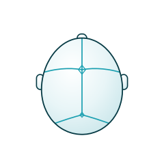
A newborn's skull is not a single bone. The frontal, side and back bones of the skull are joined by flexible lines of connective tissue called sutures. The soft areas where sutures meet are called fontanelles, or soft spots. This design does two jobs: it lets the head change shape slightly as it passes through the birth canal, and it gives the rapidly growing brain room to expand in the first years of life.
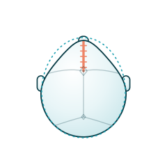
The main sutures are the sagittal suture, which runs from front to back along the top of the head; the metopic suture in the middle of the forehead; the two coronal sutures, which pass over the top of the head in front of the ears; and the two lambdoid sutures at the back of the head. The metopic suture normally closes at around 3 to 9 months, while the other sutures stay open much longer, allowing the skull to keep growing for years.
What is craniosynostosis?
Craniosynostosis means that one or more of these sutures turns to bone too early. "Synostosis" means two bones fusing together, and "cranio" refers to the skull. The skull cannot grow across the fused suture, so growth is redirected along the sutures that remain open. As a result, the head develops a recognizable shape depending on which suture is affected. Craniosynostosis occurs in roughly 1 in 2,000 to 2,500 births.
In most cases only one suture is affected and the condition appears without other health problems (non-syndromic craniosynostosis). Less often, several sutures are involved, or the condition is part of a genetic syndrome such as Apert, Crouzon, Pfeiffer, Muenke or Saethre-Chotzen syndrome (syndromic craniosynostosis). The exact cause is often unknown; in some cases genetic changes and certain factors during pregnancy may play a role.
What are the types of craniosynostosis?
Craniosynostosis is named after the suture that has fused, and each type produces its own head shape. We have detailed pages for each of the types below.
Sagittal synostosis (scaphocephaly)
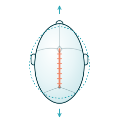
This is the most common type. The sagittal suture along the top of the head fuses early, making the head long from front to back and narrow from side to side. The forehead and the back of the head may become prominent. See our scaphocephaly (sagittal suture synostosis) page for details.
Metopic synostosis (trigonocephaly)
Here the metopic suture in the middle of the forehead fuses early. The forehead is pointed at the front and looks triangular from above, often with narrow temples and eyes that appear close together. It is important to distinguish it from a harmless metopic ridge. Details: trigonocephaly (metopic suture synostosis).
Unicoronal synostosis (anterior plagiocephaly)
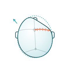
One of the coronal sutures fuses early. On the affected side the forehead flattens and the eyebrow and upper rim of the eye socket appear pulled upwards. The root of the nose and the face may deviate towards the opposite side. Details: coronal suture synostosis (anterior plagiocephaly).
Lambdoid synostosis (posterior plagiocephaly)
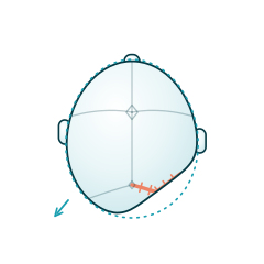
This is the rarest single-suture synostosis. One of the lambdoid sutures at the back of the head fuses early; the back of the head flattens on one side and the ear on the affected side is usually displaced downwards and backwards. It can be confused with the much more common positional flattening. Details: lambdoid suture synostosis (posterior plagiocephaly).
Bicoronal and syndromic synostosis
Both coronal sutures fuse early. The head becomes short from front to back and wide, with a tall, flattened forehead. This type is often associated with genetic syndromes, so assessment by a team including genetics, neurosurgery and craniofacial surgery is needed. Details: bicoronal (syndromic) synostosis.
What are the signs of craniosynostosis?
Signs depend on the suture involved. What they have in common is that the head takes on a shape typical of that suture, regardless of how the baby lies. Craniosynostosis is often noticed at birth or in the first months. Signs parents may notice include:
- A head that looks clearly long and narrow, triangular or asymmetrical
- A firm bony ridge you can feel along a suture line
- One-sided differences at the forehead, eyebrows or eye level
- A soft spot that closes earlier than expected
- Rarely, irritability, vomiting or developmental regression suggesting raised pressure inside the head
These signs can also appear in other, more harmless conditions. If you notice one or more of them, the first step is to talk to your pediatrician and, if needed, make an appointment with a pediatric neurosurgeon.
How does craniosynostosis differ from positional (deformational) plagiocephaly?
Most head shape differences in babies are not craniosynostosis but positional flattening. When a baby spends a lot of time lying in the same position, the back or one side of the head flattens; the sutures stay open and the skull continues to grow normally. This is called deformational plagiocephaly.
Some clues help to tell them apart. In positional flattening, the head looks like a parallelogram from above and the ear on the flat side usually moves forward. In lambdoid synostosis, the head takes on a trapezoid shape and the ear moves backwards and downwards. Positional flattening can be managed with repositioning, tummy time and, when considered necessary, a helmet, whereas craniosynostosis needs a surgical assessment. A specialist makes the final distinction.
How is craniosynostosis diagnosed?
Diagnosis begins with an examination by a pediatric neurosurgeon. The doctor looks at the head from above, the front and the back and assesses the suture lines, fontanelle, head circumference and facial symmetry, and asks about the pregnancy, birth and family history. In many cases an experienced doctor can make a strong provisional diagnosis from the examination alone.
To confirm the diagnosis or plan surgery, a three-dimensional CT scan is usually requested, with low-dose protocols generally preferred for babies. Some centers may use ultrasound for the initial assessment. If more than one suture is involved, or there are additional differences in the hands, feet or face, genetic testing may be recommended. When appropriate, an eye examination is also used to look for signs of raised pressure inside the head.
How is craniosynostosis treated?
Significant craniosynostosis is treated surgically, with the treatment planned by pediatric neurosurgery and craniofacial surgery teams. The aim is to release the area restricted by the fused suture, giving the brain room to grow, and to improve head shape. The method depends on the baby's age, the suture involved and the severity. An important point: a helmet does not treat craniosynostosis that has not been operated on, because it cannot open a fused suture.
Endoscopic surgery and the post-operative helmet
This less invasive approach is performed through small incisions and is usually chosen in the first months. The fused suture is removed as a narrow strip of bone (endoscopic strip craniectomy). Because the shape correction happens over the following months as the brain grows, a custom helmet is usually worn for several months after this operation to guide growth in the desired directions.
Open surgery (cranial vault remodeling)
This approach is usually performed somewhat later. The surgeon removes the affected skull bones, reshapes them and puts them back in place. Because most of the correction is achieved during the operation, a helmet is generally not needed routinely after open surgery, although the surgeon may recommend extra support in some cases.
The timing of surgery matters. Because the skull grows fastest during the first year, most operations are planned within this period. After treatment, head growth, eye findings and development are monitored regularly over the following years.
Where does Cranioplus fit into craniosynostosis care?
Cranioplus does not diagnose craniosynostosis or perform surgery. Our role is to design, fit and follow up custom cranial orthoses for babies whose pediatric neurosurgeon has recommended a post-operative helmet. We measure the baby's head with a contact-free, radiation-free 3D scanner, make the helmet to those measurements and adjust it for growth at check-ups. The process follows the surgical team's recommendations and is supervised by a certified orthotist and prosthetist. If a baby who has not had surgery is suspected of having craniosynostosis, we first refer the family to a pediatric neurosurgeon.
Frequently asked questions
Is craniosynostosis hereditary?
Most single-suture craniosynostosis occurs by chance, with no one else in the family affected. In syndromic types, specific gene changes play a role and can be passed from parent to child. If there are similar head shapes in the family, differences in the hands or feet, or more than one suture is involved, genetic counseling can help both with assessing the baby and with planning future pregnancies.
Can craniosynostosis improve without surgery?
A fused suture does not reopen on its own, and a helmet cannot open it either. In marked craniosynostosis the head shape usually does not improve with growth, and some children are at risk of raised pressure inside the head. However, in some very mild cases, particularly mild metopic synostosis, the doctor may recommend regular monitoring instead of surgery. The decision is made for each child together with the pediatric neurosurgeon.
At what age is craniosynostosis surgery done?
Timing depends on the method. Endoscopic surgery is usually chosen in the first months, while the skull bones are thin and the brain is growing quickly. Open surgery is mostly planned in the second half of infancy or around the first birthday. Syndromic and multi-suture synostosis may need more than one operation. That is why seeing a specialist early, if you have concerns, keeps more options open.
When is a helmet used in craniosynostosis?
A helmet is not a treatment for craniosynostosis that has not been operated on. It is used on the surgeon's recommendation, mainly after endoscopic surgery, to guide how the skull reshapes. Daily wearing time, check-up intervals and total duration are decided together by the surgical team and the orthotic team, based on the baby's age and growth rate. After open surgery, a helmet is generally not needed routinely.
This page is for general information only and does not replace a medical examination or diagnosis. Always consult a specialist about your baby's condition.
Reviewed byMemet Ateş — Prosthetist–Orthotist